
How do microbiologists see the world? What does a butterfly wing look like magnified 200 times? What is a Confocal Microscope? Britannica ImageQuest throws a new perspective on the mundane with our microscopy collection.
The following photos and images are a small sample from the Microscopy collection. Get access to the full collection plus millions of other rights-cleared educational images with a school-wide ImageQuest subscription.
Microscope image of the Lodran Meteorite
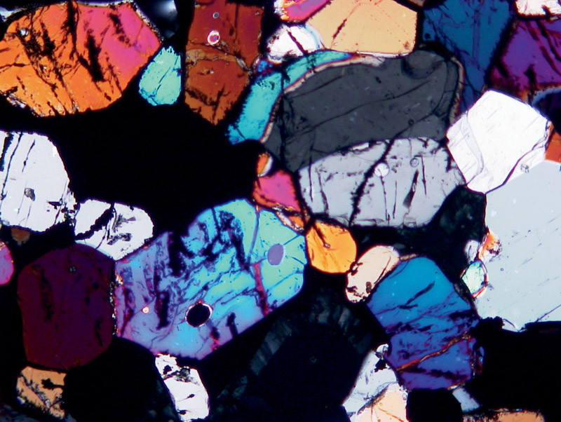
This meteorite is the type specimen of the Lodranite meteorites. The Lodranites are related to the Acaplucoites but are more course-grained. Field of view is 2.5mm across.
Fluorescence micrograph of leaf hairs
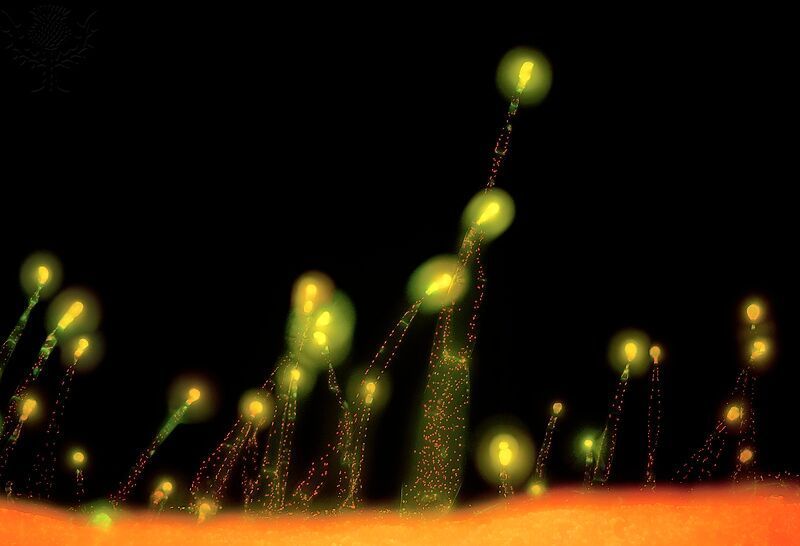
A fluorescence microscope is an optical microscope that uses fluorescence and phosphorescence instead of, or in addition to, reflection and absorption – as used by normal light microscopes.
Mycorrhiza in plant root cells
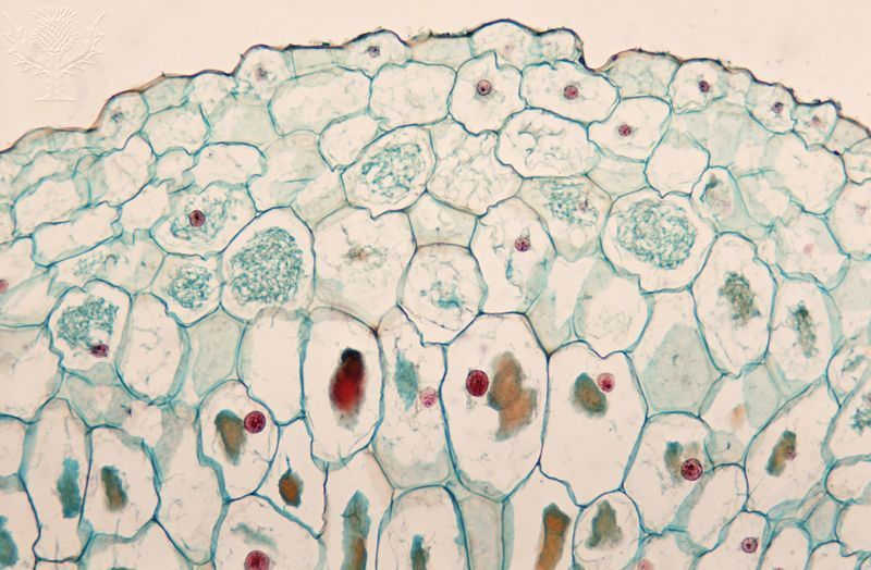
Mycorrhizza refers to an intimate and symbiotic association between the branched, tubular filaments (hyphae) of a fungus and the roots of higher plants. What can you notice about the cell walls in this image?
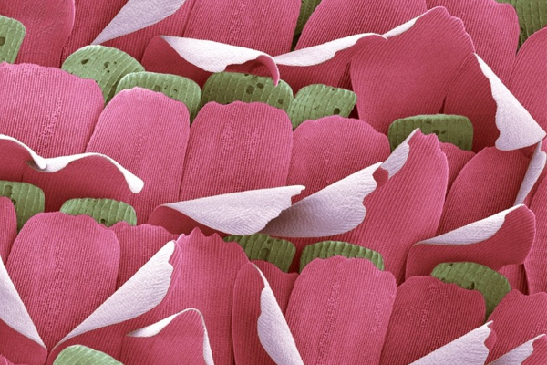
Coloured scanning electron micrograph (SEM) of the scales on a butterfly wing
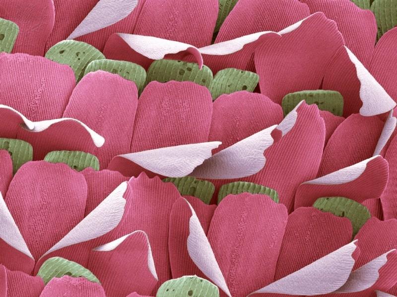
A butterfly wing is covered in many tiny scales. The scales are modified hairs made of chitin, a common substance in insect exoskeletons. The iridescence of the colours of a butterfly’s wings is produced by the diffraction of light by the microscopic structure of these scales.
Snail teeth at 1200x magnification (Scanning electron microscope)

Snails chiefly feed on plant material, but have you ever wondered how they shred and eat their food?
A composite confocal laser micrograph of freshwater green algae
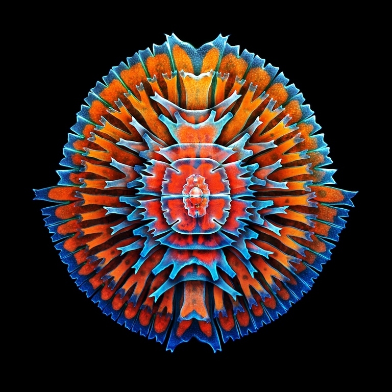
Confocal microscopy’s main feature is that only what is in focus is detected, and anything out of focus appears as black, making it particularly useful for studying the fine and three-dimensional structures of biological specimens. What differences can you notice between this image and the one of Mycorrhizza in plant root cells?
Begin your own photo adventure – Britannica ImageQuest
Search and download 3 million rights-cleared images from 63 of the world’s best collections. Get started free.
More Educator Resources
Sign up with your email for more free resources from Britannica.